
7-Micron Flat Lens Shapes Light into Optical Needle for Deeper OCT Scans
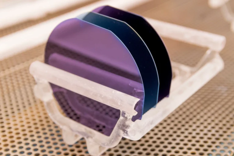
Researchers have fabricated an ultrathin flat lens that converts incoming light into an optical needle—a slender, non-diffracting beam that stays tightly focused across a long distance. Measuring roughly 7 microns in thickness, the lens could be paired with optical coherence tomography (OCT) instruments to image deeper into biological tissue without sacrificing resolution.
How the Lens Creates a Sustained Focus
Conventional lenses bend light through curved surfaces, causing beams to diverge rapidly past the focal point. The new flat lens relies on a diffractive pattern etched onto its surface to reshape the wavefront, producing a Bessel-like beam that resists spreading. Optical needles are valuable in imaging because they preserve a small spot size over an extended depth of field, overcoming the trade-off between lateral resolution and imaging depth inherent in standard Gaussian beams.
- Thickness: The lens is only about 7 microns thick, making it compatible with miniaturised optical systems.
- Beam type: The output is an optical needle with a central lobe that stays narrow for distances far beyond the Rayleigh range of a comparable focused spot.
- Fabrication: The flat design enables wafer-scale production using standard lithographic techniques, potentially reducing cost and alignment complexity.
- Integration: Because the lens is planar, it can be directly mounted onto fibre tips, endoscopes, or OCT probes.
Potential to Transform Medical Imaging
Optical coherence tomography is a non-invasive imaging technique widely used in ophthalmology, cardiology, and dermatology to capture cross-sectional views of tissue. Its resolution rivals low-power microscopy, but the imaging depth is often limited to 1–2 mm due to scattering and beam divergence. By replacing the focusing optics in an OCT system with this flat lens, clinicians could obtain sharp images from deeper layers, improving diagnostic capabilities for conditions such as skin lesions, vascular plaques, and retinal diseases.
The advance was described in Optics & Photonics News, highlighting a growing trend toward meta-optics for biomedical applications. While the lens has shown promise in proof-of-concept experiments, further work is needed to optimise efficiency and adapt the design for specific OCT wavelengths, typically around 800–1300 nm.
| Aspect | Details |
|---|---|
| Device | Ultrathin flat diffractive lens |
| Lens thickness | ~7 microns |
| Beam characteristic | Optical needle (long depth of focus, narrow waist) |
| Primary application | Deep-tissue OCT imaging |
| Key advantage | Maintains high lateral resolution over extended depth |
| Integration readiness | Prototype; compatible with existing fibre-optic OCT setups |
Future testing will assess the lens’s performance in clinical OCT configurations, with pilot studies anticipated within the next few years. If successful, the ultrathin lens could become a standard component in next-generation endoscopic and catheter-based imaging tools.
Why This Matters
Overcoming the shallow imaging depth of conventional OCT could broaden its use in cardiology and oncology, where deeper tissue penetration is essential. This flat lens approach offers a compact solution without complex optics, potentially making high-resolution deep imaging more accessible in clinical settings.
FAQ
What is an optical needle?
An optical needle is a thin beam of light that remains tightly focused over a distance much longer than a conventional focused beam. It is often generated using special diffractive or axicon lenses to overcome diffraction and maintain a small spot size.
How does this flat lens improve OCT imaging?
Standard OCT systems are limited in depth because the focused beam diverges quickly. By producing an optical needle, the flat lens keeps the beam narrow deeper into tissue, allowing sharp images at greater depths without sacrificing lateral resolution.
Why is the lens thickness significant?
At just 7 microns thick, the lens is extraordinarily compact and can be integrated directly into miniature medical devices such as endoscopes or catheters. Its flat profile also simplifies fabrication and alignment compared to bulkier conventional optics.
When could this technology become available in medical practice?
The lens is still in the research phase. Further optimisation and clinical testing are required, but prototype integration into OCT systems is expected within the next few years, potentially leading to commercial devices later this decade.
Sources
- Optics & Photonics News (optica-opn.org)
Source: Optics & Photonics News – Optics, Photonics, Physics News